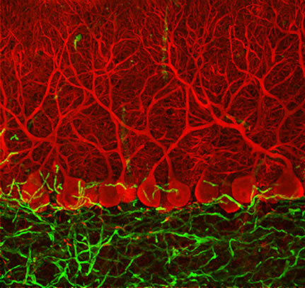
Purkinje Cells
Ludovic Collins, confocal micrograph
Wellcome Biomedical Image Awards 2006
After they get over the thrill of cutting it up, my students occasionally complain that brain tissue looks boring (somewhat like pinkish white cheese). Perhaps because the brain is so complex and beautiful in function, they expect its structure to match. And although the brain’s structure is both complex and beautiful, that’s hard to prove in an anatomy lab, because CNS neurons are small, and most have a relatively generic morphology under the light microscope.
The bipolar Purkinje cells of the cerebellar cortex are my ace in the hole for brain histology labs. They’re huge (for neurons), readily identifiable, and the copious, densely packed array of dendrites (signal-receiving processes) are clearly distinct from the single, slender axon. A single Purkinje cell receives information from hundreds of thousands of synaptic inputs.
Although Purkinje cells are not visible to the naked eye, they are quite beautiful under confocal microscopy. The stunning micrograph above depicts the Purkinje cell layer of the cerebellum in red. Each neuron has a large cell body, with a dendrite “trunk” and branches reaching upward. The effect is like a hedge (or, more accurately, rows of espaliers, since each cell’s arbor is relatively flat). The cerebellar cortex, like a large vineyard, contains row upon row like this.
The green area below the Purkinje cells is the granular layer. The single axon of each Purkinje cell reaches right through the granular layer like a taproot, sending information to the cerebellar deep nuclei, which in turn convey information to the cerebral cortex.
The Purkinje neurons, along with the rest of the cerebellum, regulate balance and coordination of movement. Injury to Purkinje cells impairs motor functions; several types of hereditary ataxias (movement disorders) are caused by the gradual death of Purkinje cells. There is a similarly named structure in the heart, the Purkinje fiber, but Purkinje fibers consist of specialized muscle cells, not neurons. Both types of cells were discovered by, and take their name from, a Czech anatomist.
Collins’ Purkinje cell micrograph is from the gallery of 2006 Wellcome Biomedical Image Awards winners, a collection of some of the most beautiful data you’re likely to see.